Tracking Stem Cells
Introduction
Although stem cells hold tremendous promise to repairing injured tissue, getting these cells to the site of injury is not always that straight-forward. Thus, investigators have relied on tracking technologies to monitor the persistence and location of stem cells after administration. Here at UC Davis, we are pioneering the use of imaging technology to monitor stem cell fate in several projects including mesenchymal stem cell mediated regeneration of the liver in dogs and acute tendon lesions in horses.
Studies
1. Evaluation of stem cell delivery strategies to the liver
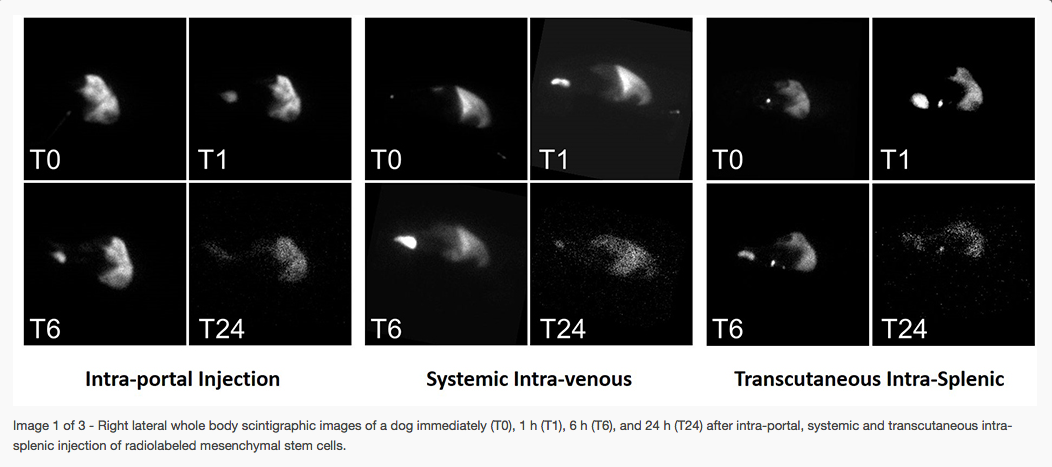
Mesenchymal stem cells (MSCs) are adult-derived, and aid in tissue regeneration by the secretion of growth factors and immunomodulatory mediators and they also stimulate and participate in vasculogenesis. MSCs can stimulate hepatic development and regeneration after hepatectomy and ischemia-reperfusion injury; they have also been used to attenuate liver fibrosis.
A technique for splenic injection using ultrasound guidance has been reported in the dog for the diagnosis of portosystemic shunt (trans-splenic portal scintigraphy). This technique could be adapted for injection of stem cells to the liver. This investigation represents a preliminary feasibility study of ultrasound-guided splenic injection of radiolabeled stem cells in comparison with portal and systemic injections regarding hepatic uptake. We hypothesized that splenic injection would be a good alternative to portal injection for hepatic delivery of stem cells and would lead to higher hepatic uptake than systemic injection. In order to track cells, MSCs were radiolabeled and imaged using scintigraphy.
Investigators: Mathieu Spriet
Results
2. Optimization of stem cell delivery to treat tendon lesions in horses
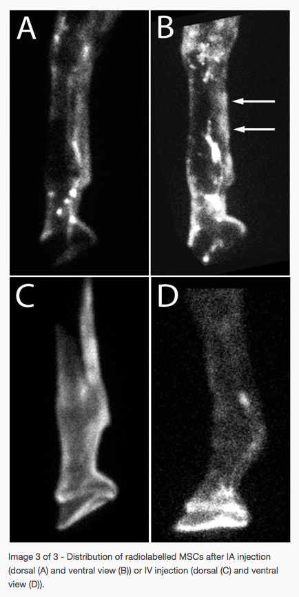
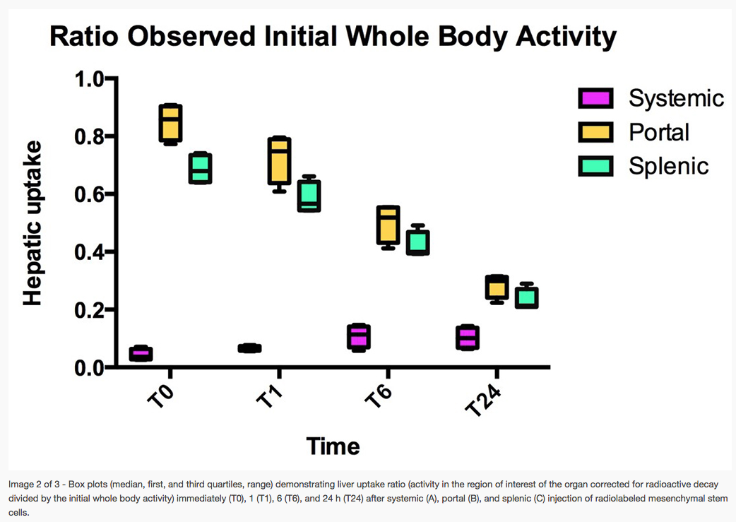
A growing body of literature suggests that MSCs are capable of mobilizing from bone marrow to home to injured tissues through chemokine and growth factor signals, which attract MSCs and facilitate migration. Our objective with this study was to evaluate whether MSCs may preferentially localize in acute tendon lesions after intravenous or intra-arterial vascular MSC administration using scintigraphic tracking of radiolabelled stem cells. This may help differentiate the most appropriate technique for MSC treatment of more subtle or inaccessible lesions.
Investigators: Mathieu Spriet
Results