Mandibular Reconstruction
Introduction
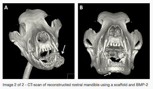
Extensive mandibular defects can be secondary to trauma, tumor resection, or other pathologic, developmental, or congenital disorders. Rostral mandibular critical-size bone defects (i.e., a defect that would not heal by bone formation during the lifetime of the animal because of the extent of the defect) result in oral misalignment and the inability to chew. Importantly, reconstruction of the rostral mandible can be challenging due to the complex anatomical geometry of this region. Particularly, the shape of the rostral mandibles in dogs resembles a sharp-angled arc, which is quite different from the geometric shape of the mandibular body and the rounded conformation in humans.
Studies
Mandibular reconstruction of critical-size defects requires rigid fixation, typically in the form of a plate and screws, and well-vascularized soft tissues. There are several strategies to fill the critical-size bone defects including autologous bone grafts, bone graft substitutes, and free-fibular flap tissue transfer. These methods, however, are not ideal as they result in donor site morbidity, are limited by graft size (especially in small dogs), and are difficult to contour. In addition, the outcome of the aforementioned may be unpredictable. Our group and others have demonstrated that a regenerative approach to reconstruction of mandibular critical-size defects in dogs using a scaffold and growth factors such as bone morphogenic protein (BMP-2) can be performed successfully and represents an excellent functional solution.


News and Media
- UC Health: Whiskey's Jaw
- Kabang on the road to recovery
- UC Davis vets treat disfigured dog
- Fox News: Mandibular reconstruction at UC Davis
- USA Today: A better jawbone
- Dogs sprout new jaw
Investigators: Boaz Arzi and Frank Verstraete
